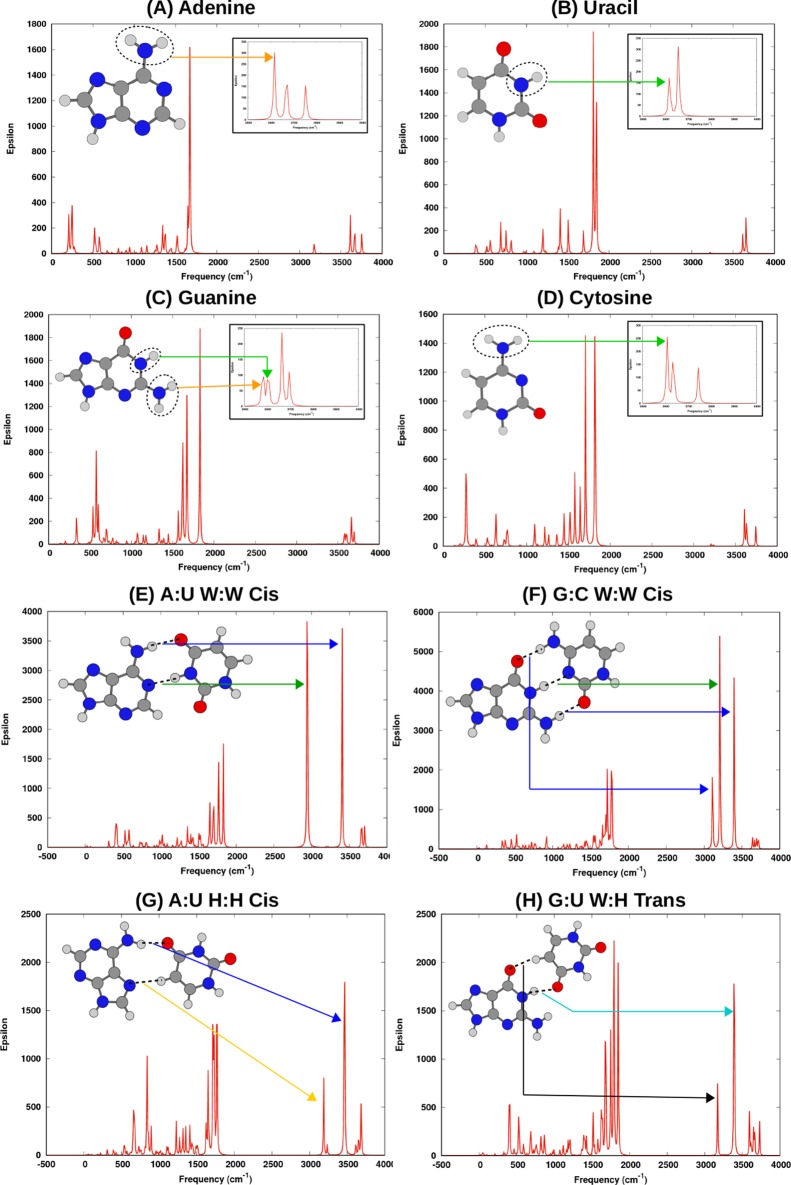
Figure 3.
IR spectra (calculated at the B3LYP-D3(BJ)/6-31+G(d,p) level) of four nucleobases (A) adenine, (B) uracil, (C) guanine, and (D) cytosine; two canonical base pairs (E) A:U W:W Cis and (F) G:C W:W Cis; and two noncanonical base pairs (G) A:U H:H Cis and (H) G:U W:H Trans. For the nucleobases, the orange arrow and the green arrow point to the frequencies corresponding to symmetric stretching of the N–H bonds of the primary amino and secondary amino groups, respectively. Interbase H-bonds of the base pairs are shown in broken line. Frequencies corresponding to symmetric stretching of the N–H (or C–H) bond in interbase H-bonds are pointed using different colored arrows for different types of H-bonds: NI–H···Oc (blue), NII–H···NIII (green), NII–H···Oc (cyan), C–H···N (orange), and C–H···O (black).

