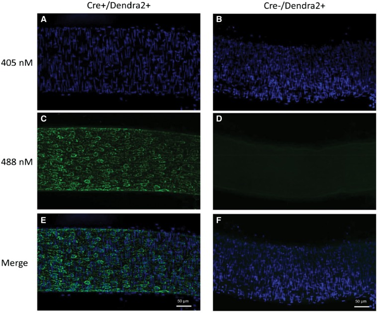
Figure 3.
Laser-scanning confocal micrographs of an intact mesenteric artery from a Cre+/Dendra2+ mouse (left; A, C, E) and a Cre−/Dendra2+ (right; B, D, F) mouse. Nuclei were stained with DAPI (A, B). Green Dendra2 fluorescence was observed only in the Cre+/Dendra2+ artery (C), while no green fluorescence was observed in the artery from the Cre−/Dendra2+ mouse (D). Merged images indicate the green fluorescence of the Dendra2 protein co-localizes with EC nuclei (E), which run parallel to the lumen of the vessel. All images were taken at ×20 magnification and are maximum projection Z-stacks. Scale bar = 50 µm. Images were taken from three consecutive mice.

