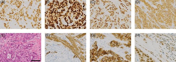
Figure 3.

Examples of typical IHC for breast tumours arising in a mutant TP53 background. Tumours are typically ER (A), PR (B) and HER2 (D) positive, show strong nuclear p53 staining (C) and are positive for markers of activated TGFβ signalling (F, αSMA; G, integrin αvβ6; H, pSMAD2/3). A corresponding H&E stain is shown in (E).
