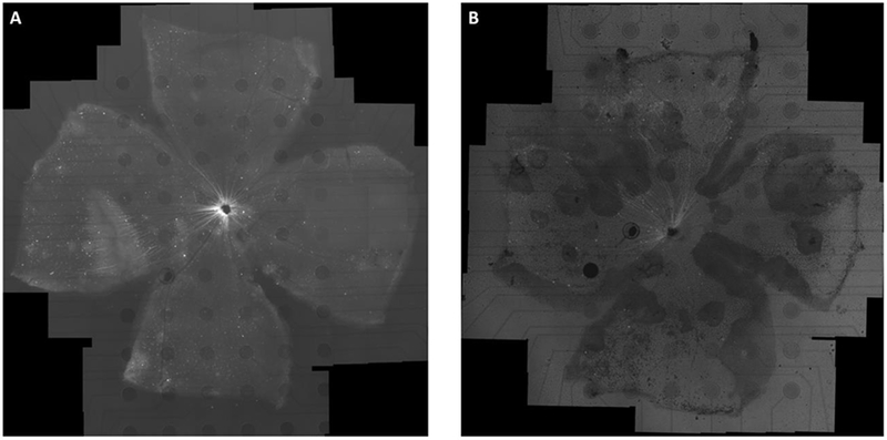
Figure 4:

Retinal whole mounts of a WT (A) and RD (B) adult mice transduced with AAV2-CAG-GCaMP6f (3 weeks post injection). The retina was mounted on a 6 × 10 rectangular MEA with a homemade retaining ring and imaged with an inverted fluorescence microscope. The baseline fluorescent image shows that GCaMP6f indicators are well expressed throughout the ganglion cell layer (GCL) for WT and most regions (with lighter but frosted texture background) for RD. The mosaic was created by stitching together 101 and 78 × 10 images respectively. The dark circles are 200μm-diameter indium tin oxide (ITO) electrodes.
