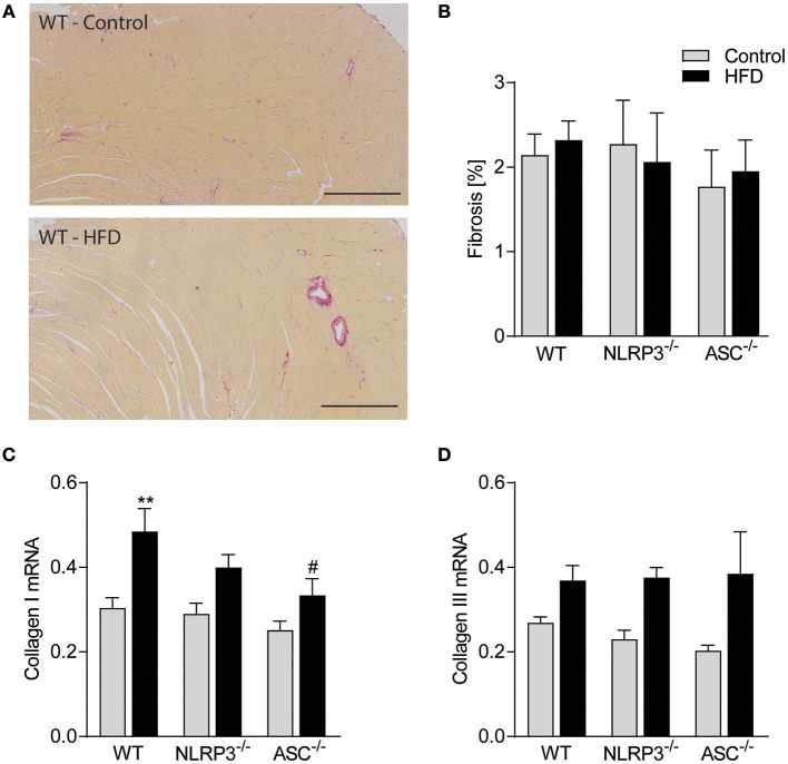
Figure 5.
Obesity-induced LV remodeling is not associated with cardiac fibrosis. WT, Nlrp3−/−, and Asc−/− (Pycard−/−) male mice were exposed to high fat diet (HFD; 60 cal% fat) or control diet for 52 weeks and cardiac fibrosis was evaluated. (A) Representative images of picrosirius red stained left ventricle (LV) from a WT mouse on control diet and HFD. Scale bar: 500 μm. (B) Quantification of picrosirius red positive areas in LV. LV expression of (C) collagen I mRNA and (D) collagen III mRNA. WT: Control, n = 10; HFD, n = 10, Nlrp3−/−: Control, n = 7; HFD, n = 7, and Asc−/− (Pycard−/−): Control, n = 7; HFD, n = 7. Data are shown as mean ± SEM. **P < 0.01 vs. control diet; #P < 0.05 vs. WT-HFD as determined by two-way ANOVA and Tukey's multiple comparisons test.

