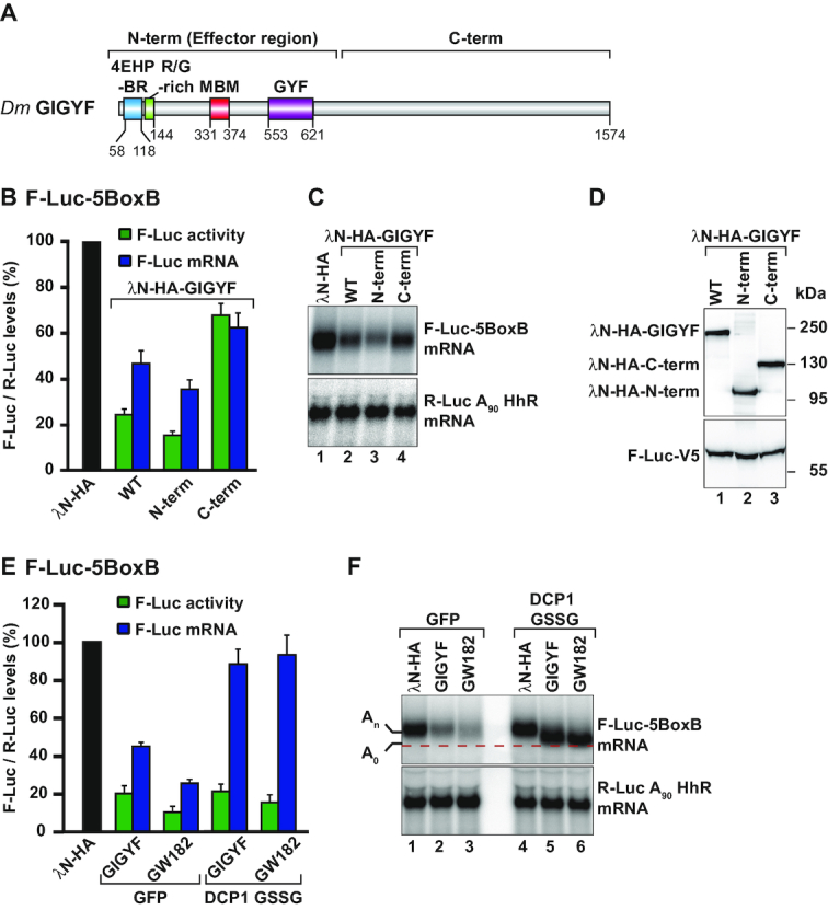
Figure 2.

Dm GIGYF represses translation and induces mRNA decay. (A) Schematic representation of the Dm GIGYF protein. The N-term contains a 4EHP-binding region (4EHP-BR), an Arg/Gly-rich sequence adjacent to the 4EHP-BR, a Me31B binding motif (MBM) and a glycine-tyrosine-phenylalanine (GYF) domain of the Smy2-type (13). The C-term does not contain any known domain and is predicted to contain primarily α-helices. The amino acid positions at the domain/motif boundaries are indicated below the protein. (B–D) Tethering assay using the F-Luc-5BoxB reporter and the indicated λN-HA-tagged proteins in S2 cells. Samples were analyzed as described in Figure 1A. Relative luciferase activity (green bars), relative reporter mRNA levels (blue bars) and representative northern and western blot images are depicted in panels B, C and D, respectively. (E, F) A tethering assay with the F-Luc-5BoxB reporter and the indicated λN-HA-tagged proteins performed in control cells (GFP-V5-expressing cells) and in cells expressing a DCP1 GSSG mutant. GW182 was used as a positive control for deadenylation-dependent mRNA decapping. Samples were analyzed as described in Figure 1A. In panel F, the red dashed line marks the position of the deadenylated (A0) F-Luc-5BoxB reporter mRNA after northern blot analysis of representative RNA samples. An indicates the position of the adenylated reporter mRNA.
