Figure 3.

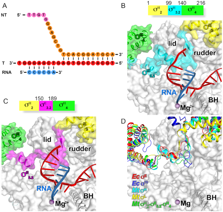
The crystal structure of Mycobacterium tuberculosis σH-RPo and σH/E-RPo. (A) The nucleic-acid scaffold used for the structure determination. (B) The schematic diagram of M. tuberculosis σH and interaction of the σ3.2-like linker of σH (σH3.2) with RNAP active-center cleft. (C) The schematic diagram of M. tuberculosis σH/E and interaction of σE3.2 with RNAP active-center cleft. (D) The domain σ3.2 of Ec σE, Ec σ70, Mtb σE, Mtb σL, Mtb σH factors follow similar path to enter and exit RNAP active-center cleft.
