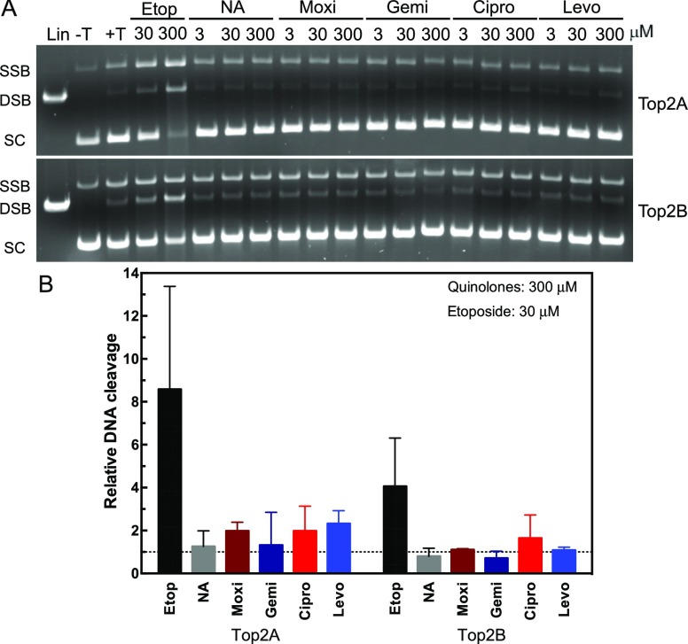
Figure 2.
Plasmid DNA cleavage by topoisomerase IIα and IIβ in the presence of quinolones. A: Plasmid DNA was incubated in the absence (−T) or presence (+T) of topoisomerase IIα (Top2A, top gel) or β (Top2B, bottom gel). Linearized plasmid (Lin) is shown in the first lane. Reactions were incubated with 30 or 300 μM etoposide (Etop) as a positive control. Reactions were performed with 3, 30, or 300 μM of nalidixic acid (NA), moxifloxacin (Moxi), gemifloxacin (Gemi), ciprofloxacin (Cipro), or levofloxacin (Levo). Positions of SSB, DSB, and supercoiled (SC) DNA are denoted at left. Representative gels are shown, and experiments were performed at least three times. B: quantification of double-strand DNA cleavage relative to cleavage in the absence of quinolones is shown for both topoisomerase II isoforms. As a control, etoposide at 30 μM is shown compared with 300 μM of the quinolones. Dotted line represents the level of cleavage in the presence of dimethyl sulfoxide (DMSO). Error bars represent the standard deviation of three or more independent experiments.

