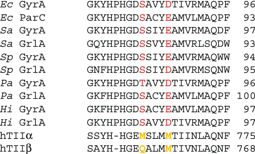
Figure 5.
Alignment of selected enzyme sequence at the region known to bind to quinolones. Highlighted in red are the positions of the Ser and Asp/Glu residues. Shown are sequences for DNA Gyrase GyrA and topoisomerase IV ParC/GrlA subunits from Escherichia coli (Ec), Staphylococcus aureus (Sa), Streptococcus pneumonia (Sp), Pseudomonas aeruginosa (Pa), and Haemophilus influenza (Hi). Also shown are sequences for Homo sapiens topoisomerase II (hTIIα and hTIIβ), which lack the residues involved in the quinolone salt bridge.

