Figure 2.

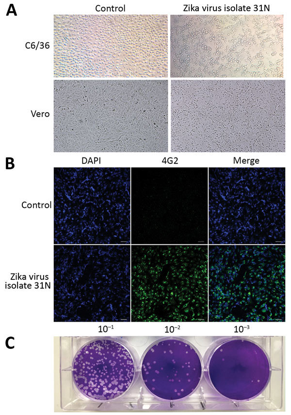
Phenotypic analysis of Zika virus isolate 31N from an Aedes aegypti larval pool, Jojutla, Morelos, Mexico. A) Cytopathic effect of the Zika virus isolate 31N in C6/36 and Vero cells. The left panel shows mock infected cells. Original magnification ×20. B) Infected Vero cells with Zika virus isolate 31N at a multiplicity of infection of 0.1 and mock infected cells. Nuclei are stained in blue (DAPI), and the envelope protein is stained in green (4G2). Original magnification ×20. C) Plaque assay of Zika virus isolate 31N in Vero cells. Serial decimal dilutions of Zika virus isolate 31N are depicted.
