Figure 1.

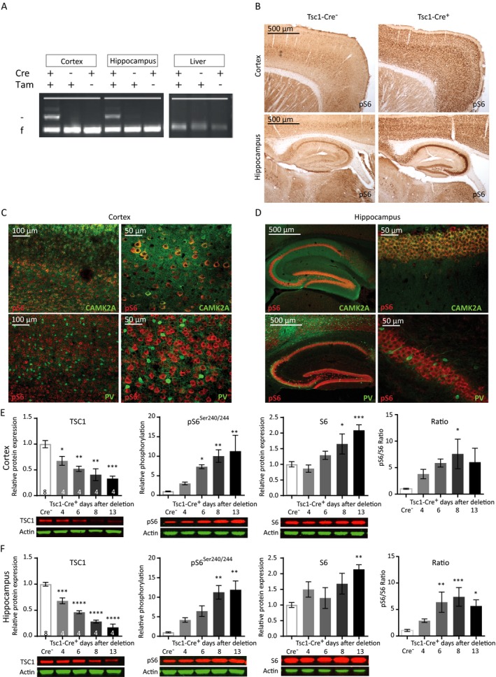
Cre‐mediated Tsc1 gene deletion in CAMK2A‐expressing neurons induces hyperactivation of the mTORC1 pathway. (A) Tsc1 PCR analysis of DNA isolated from the cortex, hippocampus, and liver after four tamoxifen (Tam) injections, inducing efficient and brain‐specific Tsc1 gene deletion of mice sacrificed at day 11. The higher band indicated with ‘–’ represents the knock‐out allele; the lower band ‘f’ represents the floxed (nondeleted) allele. (B) Immunohistochemistry (DAB staining) on sagittal sections of an adult Tsc1‐Cre+ and Tsc1‐Cre– mouse, 13 days after gene deletion stained for pS6Ser240/244. Increased levels of pS6Ser240/244 in tamoxifen injected Tsc1‐Cre+ mice are observed. (C and D) Fluorescent immunohistochemistry on coronal sections of Tsc1‐Cre+ for pS6Ser240/244 shows high colocalization with CAMK2A (green) but not with parvalbumin (PV; red), in the cortex and hippocampus. (E and F) Tsc1‐Cre+ mice show TSC1 reduction and increased levels of S6 and pS6Ser240/244 starting at day 6 after gene deletion in the cortex and hippocampus. Western blot data are presented as means with error bars representing SEM. Sample sizes are indicated in the figures with n the number of mice used. A one‐way ANOVA was used for statistical analysis with a Dunnett’s post hoc test. Tsc1‐Cre+ mice were used as control group. *P < 0.05, **P < 0.01, ***P < 0.001, ****P < 0.0001.
