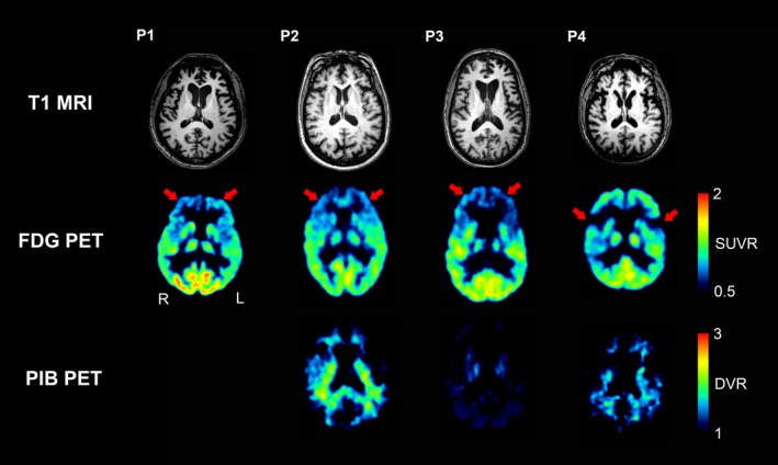
Figure 2.

Brain magnetic resonance (MR), 18F‐FDG PET, and 11C‐PIB PET images of patients with frontotemporal lobar degeneration (FTLD). Patients 1, 2, and 3, who had behavioral variant frontotemporal dementia (P1, P2, and P3), showed atrophy and hypometabolism in the bilateral anterior frontal cortices on MRI and 18F‐FDG PET, respectively. Patient 4, who had progressive nonfluent aphasia (P4), showed bilateral perisylvian atrophy and hypometabolism on MRI and 18F‐FDG PET, respectively. 11C‐PIB uptake was not increased in these areas or other gray matter areas for any of the patients. 11C‐PIB PET imaging was not available for Patient 1. Abbreviations: FDG, fluorodeoxyglucose; PIB, Pittsburgh Compound B.
