
Figure 2.
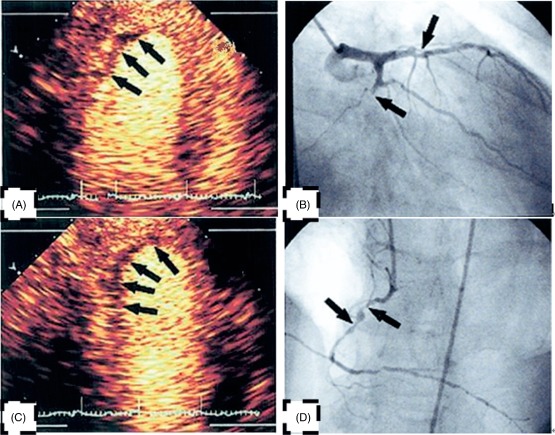
(A) Signal intensity (SI) of mid inferior wall and apex inferior wall decreased conspicuously. (B) The left circumflex occlusion corresponds to (A). (C) SI of mid posterior wall and apex posterior wall decreased conspicuously. (D) The right coronary artery occlusion corresponds to (C).