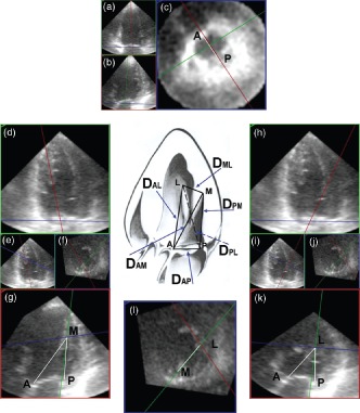
Figure 1.

Measurement of the mitral tetrahedron. First, the maximal left ventricular (LV) contraction was defined as the end‐systolic phase. An anteroposterior plane (red frame) that passed through the anterior and posterior annulus was adjusted to parallel the LV axis (part a). Another plane (green frame) perpendicular to the anteroposterior plane was also adjusted to parallel the LV axis (part b). Then, a horizontal plane (blue frame) was accommodated to the mitral ring by the assistance of these 2 planes (part c). The distance between the anterior and posterior annulus (DAP) was measured, and the long‐axis of mitral annulus was also obtained. The mitral annular plane (part c) was annotated to form the reference plane for tilting the anteroposterior plane. Second, the blue frame was moved to the level of the roots of the papillary muscles and intersected with the green frame. These 2 frames were moved carefully to define the exact point of the roots of papillary muscles (part l). The blue frame at this time was annotated to be the papillary muscle roots plane. The anteroposterior plane (red frame) was tilted to the root of medial papillary muscle using a reference hinge intersecting by the mitral annular plane and the anteroposterior plane (part d). The exact intersection of anteroposterior plane with the root of the medial papillary muscle was assisted by the green frame and the papillary muscle roots plane (parts e and f). On the anteroposterior plane imaging (part g), the intersecting point of the blue and green line was the root of medial papillary muscle (M) (part g), and the distance between the root of medial papillary muscle and the anterior annulus (DAM) and the posterior annulus (DPM) could be measured. By the same way, the distance between the root of lateral papillary muscle and the anterior annulus (DAL) and the posterior annulus (DPL) could be measured after tilting the anteroposterior plane to the root of lateral papillary muscle (parts h–k). Finally, the interpapillary muscle roots distance (DML) could be measured on the papillary muscle roots plane (part l). Abbreviations: M, the root of medial papillary muscle; L, the root of lateral papillary muscle; A, the anterior annulus; P, the posterior annulus.
