Figure 1.

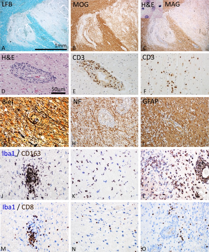
Pathological features of inflammatory demyelinating brain lesions in index patient A. (A–C) Focal plaques of demyelination with loss of myelin in Luxol fast blue myelin stain (A), partial preservation of myelin oligodendrocyte glycoprotein (MOG) reactivity in particular in initial lesion stages (B), but complete loss of myelin associated glycoprotein (MAG; C) and apoptotic nuclear changes of oligodendrocytes (C, insert). Magnification bar is applicable for (A–C). (D–F) The demyelinated lesions occur on the background of profound brain inflammation, reflected by perivascular cuffs seen in hematoxylin/eosin staining (D). The inflammatory reaction dominantly contains CD3+ T‐lymphocytes, which are present within the perivascular cuffs (E) and diffusely dispersed in the lesion parenchyme (F). (G–I) Characteristic features of the lesions are preservation of axons within the lesions, shown by Bielschowsky silver impregnation (Biel) (G) and immunohistochemistry for neurofilament protein (NF) (H) and the pronounced astrocytic gliosis, seen after staining for glial fibrillary acidic protein (GFAP, I). (J–L) Double staining for the pan microglia/macrophage marker Iba‐1 (blue) and the macrophage activation antigen CD163 (brown) in the periplaque white matter with microglia clusters (J), in initial lesions with diffuse microglia activation (K) and in the demyelinated lesion center with profound macrophages infiltration and activation (L). Only a subset of microglia/macrophages also expresses the activation marker CD163. (M–O) Double staining for the pan microglia/macrophage marker Iba 1 with CD8 in the periplaque white matter (M), the initial lesion (N), and the active demyelinated lesion (O). In all lesion stages CD8 cells can be seen, which are in close apposition with microglia/macrophages. Magnification bar in (D) applicable for (D–O).
