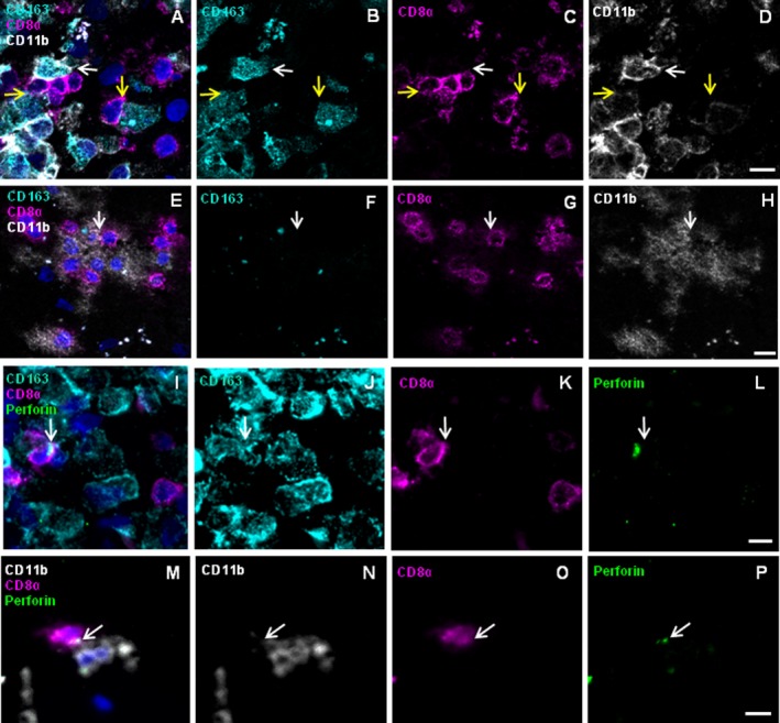
Figure 4.

Immunohistochemistry reveals CD8+ T cells juxtaposed to CD163+ and CD11b+ MPs showing vectorial perforin orientation in patient A. Frozen sections of brain lesions of patient A were stained for the markers CD8 (magenta), CD163 (cyan), CD11b (white), perforin (green), and nuclei (DAPI, dark blue). Rows 1 and 2 show the four‐color overlay of CD163, CD11b and CD8 (A and E) and single color staining for each of the antigens CD163 (B and F), CD8 (C and G), and CD11b (D and H). CD8+ cells were detected in close physical contact with CD163+ CD11bhigh+ as well as with CD163+ CD11bweak+ cells (A–D) as indicated by white and yellow arrows, respectively. Furthermore, CD8+ cells contacted CD163− CD11b+ cells (E–H). Rows 3 and 4 show the four‐color overlay of CD8 and perforin with either CD163 (I) or CD11b (M) and single color staining for each of the antigens CD163 (J), CD11b (N), CD8 (K and O), and perforin (L and P). Arrows indicate accumulations of perforin‐containing granules. (I–L) One of several CD8+ cells orients big perforin granules toward a CD163+ cell. The CD8+ cell has a round shape and the granules are in the cytosol between the nucleus and the cytoplasmic membrane. (M–P) CD8+ cell orients perforin granules toward a CD11b+ cell. Scale bars 10 μm.
