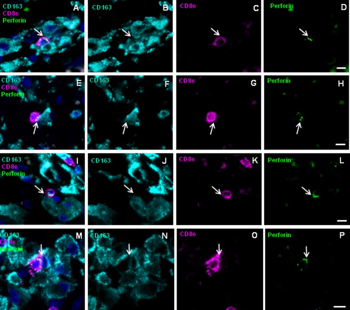
Figure 6.

Vectorial perforin orientation of CD8+ cells toward CD163+ MPs in brain lesions of other patients with MS. FFPE fixed tissue sections were stained for the markers CD163 (turquois), CD8 (magenta), perforin (green), and nuclei (DAPI, dark blue). Shown are a perivascular region of AMS patient MS11 (A–D), a parenchymal region of MS11 (E–H), a perivascular region of AMS patient MS10 (I–L), and a perivascular region of SPMS patient MS25 (M–P) The four‐color overlay (A, E, I, and M) and single color staining for each of the antigens CD163 (B, F, J, and N), CD8 (C, G, K, and O) and perforin (D, H, L, and P) is shown. Arrows indicate accumulations of perforin‐containing granules toward CD163+ cell. Scale bars 10 μm.
