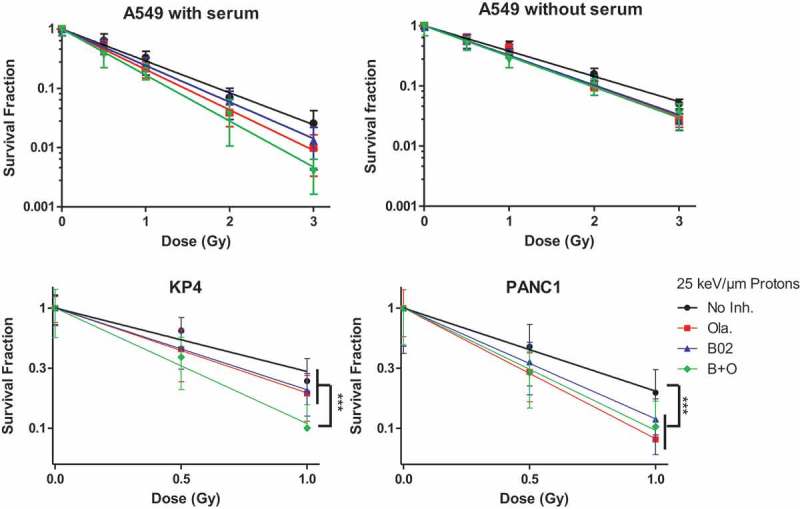
Figure 6.

Survival fraction. A549, KP4 and PANC1 cells were exposed to 225 kV X-rays or 25 keV/µm broad proton beam at 2 Gy/min. 3 to 4 h before irradiation the cells were incubated in medium without serum (A549) or with serum (A549, KP4, PANC1). The survival fraction was determined using clonogenic assays. Cells were incubated for a total duration of 24 h with: No Inhibitor (black dot); 0.5µM Olaparib (red square); 10 µM B02 (blue up triangle), 10 µM B02 0.5 µM + Olaparib (green down triangle) then in control culture medium for 11 days. At least three independent experiments were performed, and data are presented as mean ± 1 SD. The data have been fitted using Linear model. One way ANOVA (Kruskal–Wallis) statistical analyses were performed (p > 0.05: ns; p < 0.05: *; p < 0.01: **; p < 0.001: ***).
