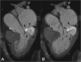
Figure 4.

Computed tomography reconstructions parallel to the long axis of the left ventricle during midsystole (A) and mid‐diastole (B) demonstrate the centrally hyperdense, peripherally calcified mass (arrowheads) located in the posterior mitral annulus and attached to the posterior leaflet. Reprinted with permission from Elsevier.
