Figure 4.
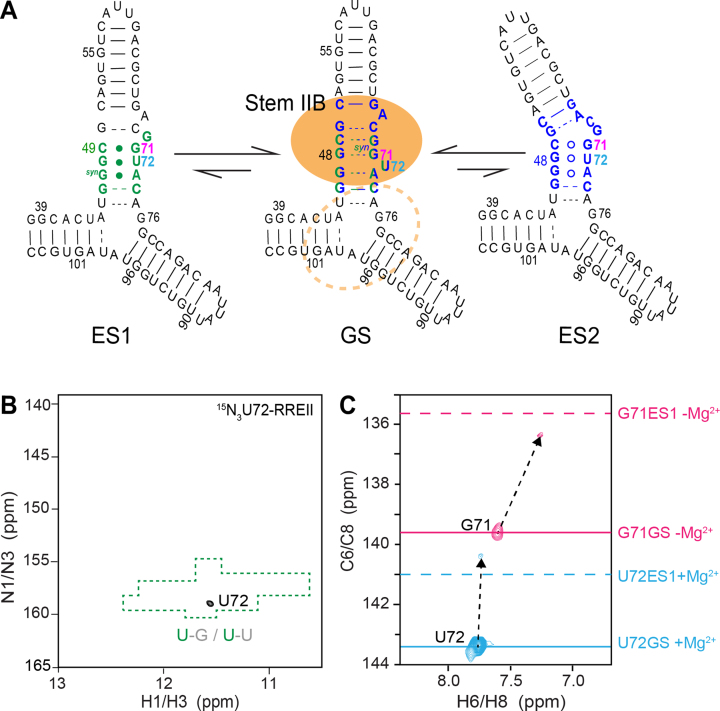
Selective labeling strategy to probe conformational exchange in RREII. (A) Proposed secondary structure for ES1 and ES2 in RREII based on ESs observed in RREIIB. Nucleotides that experience exchange due to ES1 and ES2 are colored green and blue, respectively. Rev primary and secondary binding sites are highlighted using a filled and dashed orange circle, respectively. (B) 2D 1H,15N-HSQC spectrum of site-specifically labeled 15N3-U72-RREII at 25°C in 3 mM Mg2+ showing a single imino resonance at the characteristic chemical shift region (highlighted in green) expected for a G-U wobble. (C) 2D 1H,13C-HSQC spectra of site-specifically labeled 13C6-U72-RREII (pink) and 13C8-G71-RREII (cyan) at 25°C showing the appearance of major resonances consistent with the GS in RREIIB (solid line) and minor resonances consistent with ES1 in RREIIB (dashed line). Spectra for 13C8-G71-RREII were measured in the absence of Mg2+. The sample conditions were 0.2–0.3 mM RREII in 15 mM sodium phosphate, 25 mM NaCl, 0.1 mM EDTA, pH 6.4 with or without 3 mM MgCl2.

