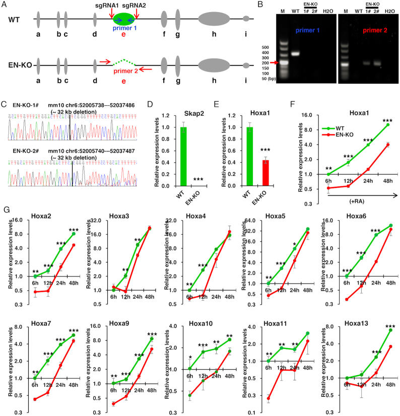
Figure 2.
e-site enhancer knockout inhibits RA-induced Hoxa1 expression. (A) Schematic showing CRISPR/Cas9-mediated deletion of the e-site enhancer (green) using two sgRNAs. Indicated primers were used to distinguish EN-KO (enhancer knockout) from wild-type (WT) clones. (B) Validation of knockout lines by genomic DNA PCR. Shown are images from representative clones. (C) DNA sequencing of two EN-KO clones (#1 and #2) using primer 1. (D and E) Skap2 and Hoxa1 mRNA levels in undifferentiation ESCs, as measured by qRT–PCR and normalized to Gapdh levels. (F) Hoxa1 mRNA levels in EN-KO and WT cells over the course of RA-induced ESCs differentiation. mRNA levels were measured by qRT–PCR and normalized to Gapdh levels. (G) Hoxa2–a13 mRNA levels were also measured by qRT–PCR and normalized to Gapdh levels in EN-KO and WT cells following RA treatment. Data are represented as mean values ± s.d. Indicated significance is based on Student's t-test (*P < 0.05, **P < 0.01, ***P < 0.001). In (D–G), n = 3 or 6, including 1 WT, 2 EN-KO cell lines (EN-KO-1# and EN-KO-2#) and three technical replicates per cell line. M: DNA Marker.

