Fig. 4.

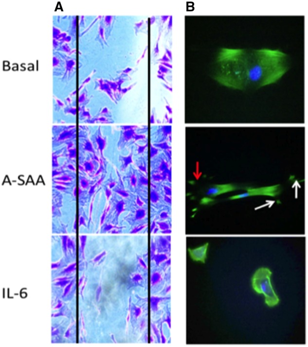
IL-6 does not promote myofibroblast migration nor cytoskeleton rearrangement
(A) Representative micrography in basal and stimulated conditions, showing myofibroblasts repopulating the wound in response to A-SAA but clear wound margins still present after exposure to IL-6. (B) In basal and IL-6-stimulated conditions, intact F-actin fibres are present. However, after treatment with A-SAA, filiopodia formation (white arrows) and membrane ruffling (red arrow) are evident.
