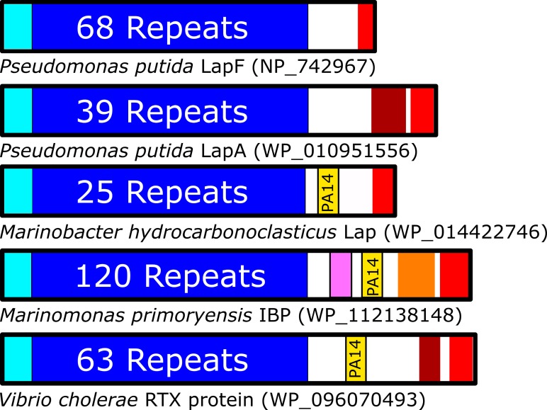
Fig 1. Organization of adhesins with and without PA14 domains.
Domain architectures are shown for several RTX adhesins, oriented with N termini on the left. Colour scheme: cyan = bacterial membrane anchoring domains, blue = region of tandem repeat domains, yellow = PA14 domain, burgundy = von Willebrand Factor A-like domain, pink = Vibrio-like peptide-binding domain, orange = ice-binding domain, red = Type 1 Secretion Signal and RTX repeats, white = regions of unknown/unpredicted composition.

