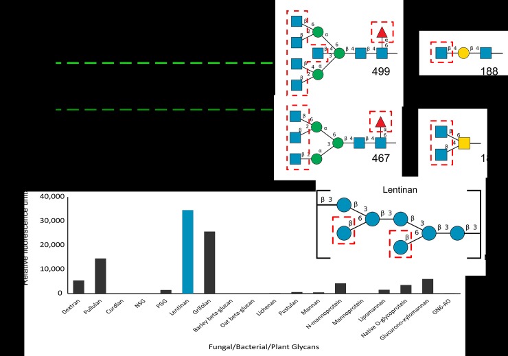
Fig 10. Binding of MhPA14 to Glycan arrays.
A) Fluorescent measurements of an array of 585 glycans of varying complexity following incubation with GFP_MhPA14 (200 μg/mL). The fluorescence measured for each glycan is an average of four replicate spots. The four glycan spots that fluoresced above 8000 RFU are labelled, and their structures are presented on the right. Blue squares = N-acetylglucosamine, blue circles = glucose, yellow squares = N-acetylgalactosamine, yellow circles = galactose, green circles = mannose, red triangle = fucose. Terminal sugars proposed to be strong-binders via the competitive assay are outlined in red. B) Fluorescent measurements from a second array, containing eighteen glycans extracted from biological sources following incubation with GFP_MhPA14 (50 ug/mL) and detected through anti-GFP antibody. The fluorescence measured for each glycan is an average of two replicate spots. The highest fluorescing glycan is coloured blue, and its repeating structure is shown on the right using the same colour scheme as in A).

