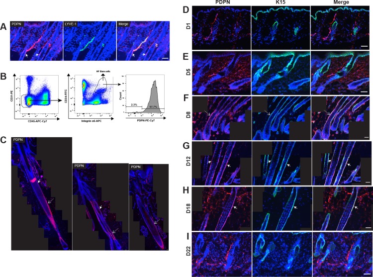
Fig 1. PDPN is expressed in the HF keratinocyte region and the stem cell area during the late anagen but not the telogen phase.
(A) Immunofluorescence staining of 10-μm frozen sections of back skin (anagen growth phase, C57BL/6J female mice at day 12 after depilation) for PDPN (red) and LYVE-1 (lymphatic marker, green). Nuclear staining with Hoechst 33342 (blue). (B) FACS gating strategy for sorting HF stem cells. Left panel: Skin-derived cell suspension pre-gated for living (7AAD-) singlets. Middle panel: CD45- CD31- CD34+ integrin α6+ cells were considered as HF-stem cells. Right panel:PDPN+ cells were dectected in HF stem cells. (C) Immunofluorescence stainings of 10-μm paraffin sections for PDPN (red) in human scalp tissue. Nuclear staining with Hoechst 33342 (blue). (D-I) After depilation-induced HF regeneration in C57BL/6J female mice (n = 4 each group), back skin samples were obtained at days 1 (early-anagen phase), 5 (mid-anagen phase), 8 (late-anagen phase), 12 (late-anagen phase), 18 (catagen phase), and 22 (telogen phase). Double immunofluorescence stainings of 10-μm frozen sections of back skin for PDPN (red) and K15 (green). Nuclear staining with Hoechst 33342 (blue). (C, F, G, and H) Tiled images from each immunofluorescence image were created to visualize large fields. White arrows indicate the bulge area. Dashed arrows indicate the HF keratinocyte region. Scale bars: 50 μm.

