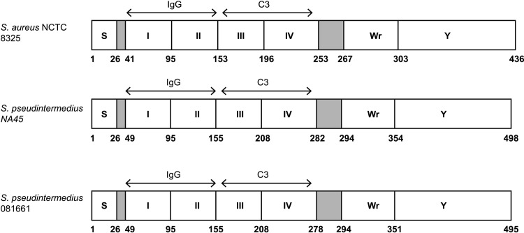
Fig 1. Schematic representation of S.pseudintermedius Sbi domains.
The predicted structures of S. pseudintermedius Sbi proteins from strains NA45 and 081661 are depicted in comparison with S.aureus NCTC 8325 [11]. These include the signal peptides (S), N-terminal IgG-binding domains I and II (corresponding to S. aureus regions D1 and D2) and domains III and IV that correspond to complement C3 binding domains D3 and D4 of S. aureus [30]. These are followed by the Wr domain which is rich in proline and the Y domain that is tyrosine rich and likely involved in membrane binding [30]. Numbers correspond to amino acid residues.

