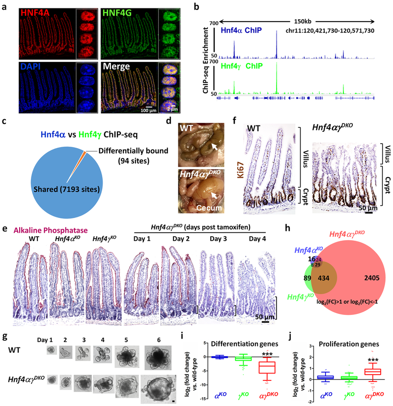
Figure 1. HNF4A and HNF4G are redundantly required to drive intestinal differentiation.
a, Co-localization of HNF4A and HNF4G in intestinal epithelial cells by immunofluorescence confocal microscopy (n = 3 biologically independent mice). The high-resolution images are of villus epithelial cell nuclei as shown in the boxed insets. ChIP-seq reveals strikingly similar binding profiles of HNF4A and HNF4G in the duodenal epithelial cells. b, Example of ChIP-seq tracks. c, DiffBind analysis (FDR < 0.05, n = 2 biologically independent mice) shows that 7,193 sites are shared and only 94 sites are differentially bound by HNF4A and HNF4G. Statistical tests were embedded in DiffBind. d, Necropsy reveals a distended and fluid-filled intestine in the Hnf4αγDKO mouse after 4 days of tamoxifen injection (n = 10 independent experiments). e, Alkaline Phosphatase staining (differentiation marker, pink color, n = 4 biologically independent mice). f, Immunostaining of Ki67 (proliferation marker, brown nuclei). Brackets show elongating crypts in the double mutants (n = 4 biologically independent mice). The proliferation zone is defined by the distribution of Ki67-positive cells. g, In contrast to WT, Hnf4αγDKO organoids show a spherical morphology and fewer lumenal contents, consistent with a failure to differentiate (n = 6 independent experiments). Scale bar, 50 μm. h, Only Hnf4αγDKO mice show a striking alteration of gene expression as evidenced by RNA-seq (n = 3 biologically independent mice). Venn diagram shows the numbers of genes in HNF4 single and double mutants with log2 fold-change > 1 or < −1, FDR < 0.05. Statistical tests were embedded in Cuffdiff. Differentiation genes (i) are reduced while proliferation genes (j) are elevated in the Hnf4αγDKO mice. The middle line represents the median; whiskers represent the 10th and 90th percentile. Post-hoc Dunn’s test was applied following a Kruskal-Wallis test at P < 0.001 ***; n = 3 biologically independent mice).

