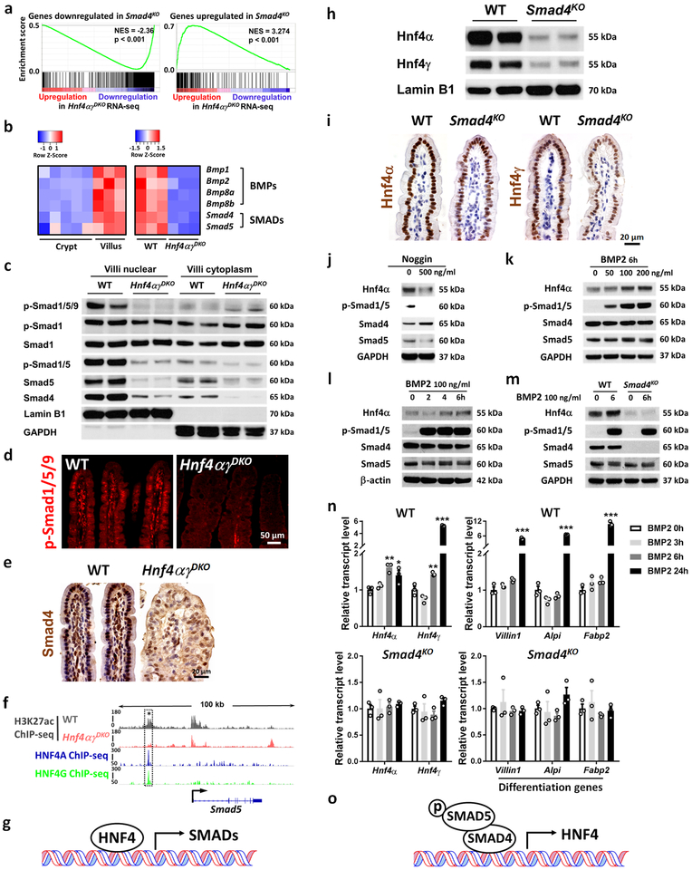
Figure 3. HNF4 and BMP/SMAD reinforce each other’s expression.
a-g, HNF4 binds and activates core components of the BMP/SMAD signaling pathways. a, GSEA reveals that genes downregulated or upregulated upon SMAD4 loss strongly correlate with genes of reduced expression or increased expression in Hnf4αγDKO, respectively (Kolmogorov-Smirnov test, one-sided for positive and negative enrichment scores, P < 0.001, n = 3 biologically independent mice). NES: normalized enrichment score. Heatmap (b) displays that RNA-seq expression levels of BMPs/SMADs are highly expressed in villus compared to the crypt and are significantly reduced in the Hnf4αγDKO intestinal epithelium compared to the WT littermate controls (n = 3 biologically independent mice, FDR < 0.001). Statistical tests were embedded in Cuffdiff. This is also evidenced by (c) western blot (n = 2 independent experiments and 2 biologically independent mice for each experiment). Immunofluorescent staining of p-SMAD1/5/9 (d) and immunohistochemistry staining of SMAD4 (e) also show reduced levels upon HNF4 loss (representative of 3 biologically independent mice). f, ChIP-seq tracks (n = 2 biologically independent mice) show that HNF4 factors bind to Smad5 and loss of HNF4 results in reduced H3K27ac signal (see dashed rectangles). Reciprocally, HNF4 levels are dependent upon BMP/SMAD4. h-o, BMP/SMAD signaling promotes HNF4 expression. Western blot (n = 2 independent experiments and 2 biologically independent mice for each experiment) (h) and immunostaining (n = 3 biologically independent mice) (i) show that SMAD4 knockout (4 days after tamoxifen injection) can cause significant downregulation of nuclear protein levels of Hnf4α and Hnf4γ in the villi. Western blot of the primary WT intestinal organoids in presence of (j) Noggin treatment (BMP antagonist, 72 h, n = 2 independent experiments), (k) 6 h treatment of different dosages of BMP2 (n = 3 independent experiments), and (l) different durations of BMP2 treatment (n = 3 independent experiments). m, BMP2 can induce Hnf4α expression in the primary WT intestinal organoids, but this effect is abolished in the absence of Smad4 (n = 2 independent experiments). Uncropped western blots are shown in Supplementary Fig. 11. qRT-PCR (n) shows that BMP2 (100 ng/ml) promotes Hnf4 and differentiation gene expression in the primary WT intestinal organoids but not in the Smad4KO organoids. All the qPCR data are presented as mean ± SEM (n = 3 independent organoid cultures). Transcript levels relative to BMP 0 h, and statistical comparisons were performed using one-way ANOVA followed by Dunnett’s post test at P < 0.001 ***, P < 0.01 ** and P < 0.05 *. All the primary organoids were harvested at Day 6 after seeding.

