
Figure 2. Pulvinar Lesion-Induced Gain Imbalance Produces Asymmetric Attentional Deficits.
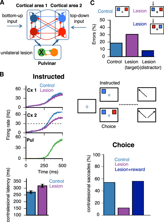
(A) Schematic as in Figure 1, where external inputs are labeled as either bottom-up (sensory) or top-down (internal), with pulvinar excitability λ = 230 Hz/nA. A unilateral lesion to the medial pulvinar is shown that affects the left visual field. Topography thus corresponds to visual and not anatomical space. (B) Visuospatial task based on Wilke et al. (2013), where a subject must make a saccade toward a visual target after a delay period (instructed) or select one of two simultaneously presented visual targets on opposite sides of the visual field (choice). In the instructed task, saccade latencies toward the contralesional field are larger than in controls. In the choice task, the proportion of saccades to the contralesional field is reduced compared to controls but ameliorated with the addition of reward (Wilke et al., 2013). Data are represented as mean ± SD. (C) Visuospatial task modeled after Desimone et al. (1990), where a subject must attend to and select a target (blue) that was flashed at the same position as a cue presented during fixation. A distractor (red) is presented simultaneously in the opposite hemifield. Simulations are performed for control and unilateral lesion of the lateral pulvinar. Black arrows point to the affected visual hemifield, and two conditions can be distinguished: either the target (magenta) or the distractor (dark blue) lies within the affected hemifield. Error rates are shown below.