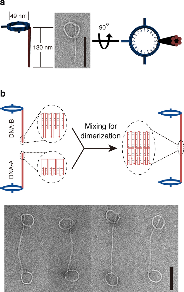
Figure 1. DNA nanostructures designed for this study.
a, Cartoon models and a negative-staining TEM image of monomeric DNA nanostructure comprising a ring connected to a rod. b, Schematics of DNA nanostructure dimerization. Diagrams in the dashed ovals illustrate the dimerization mediated by DNA rods with complementary sticky ends. Negative-stain TEM images of DNA-origami dimers (two rings joint by a pillar) are shown at the bottom. Scale bars: 100 nm. The experiments were repeated independently three times with similar results.

