
Figure 1.
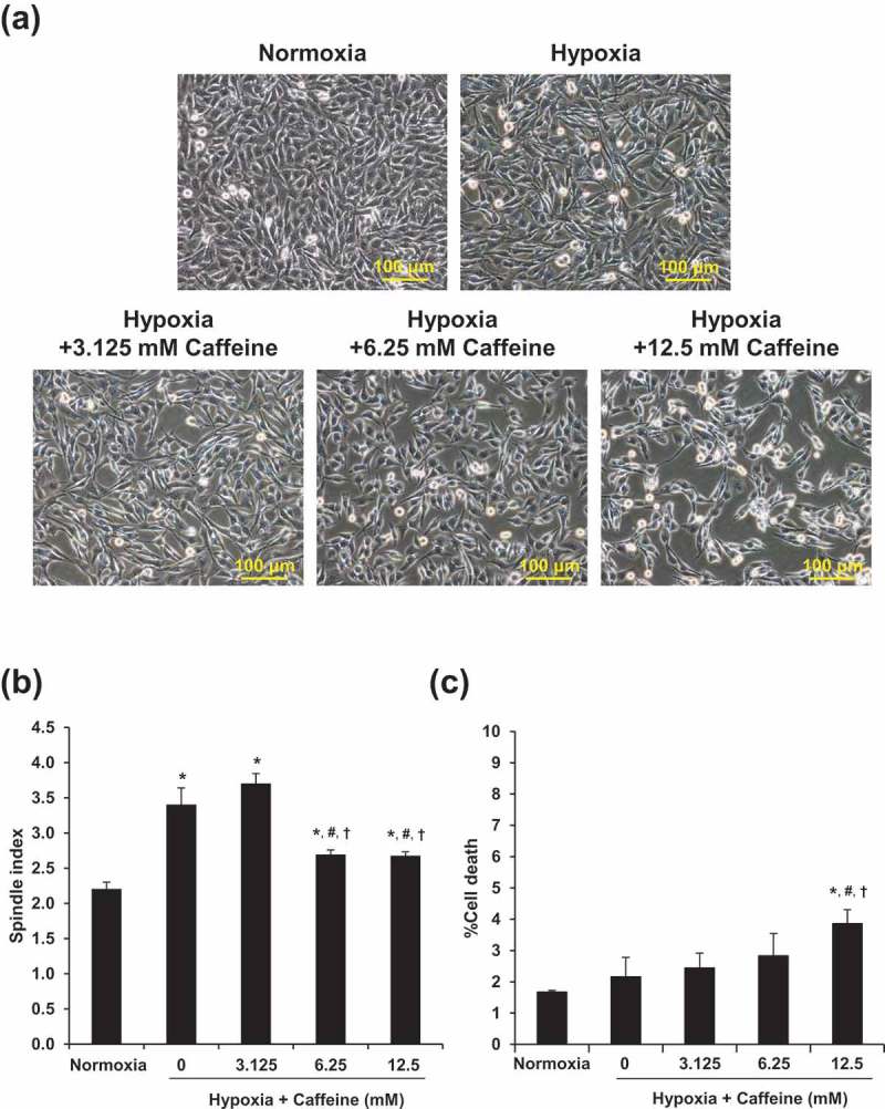
Validation of the study model. (a): Cell morphology was investigated under an inverted phase-contrast microscope (original magnification power = 200X for all panels). (b): Spindle index. (c): Percentage of cell death. Each bar represents the mean ± SEM of the data derived from three independent biological replicates. * p < 0.05 vs. control (normoxia); # p < 0.05 vs. hypoxia; †p < 0.05 vs. hypoxia + 3.125 mM caffeine.