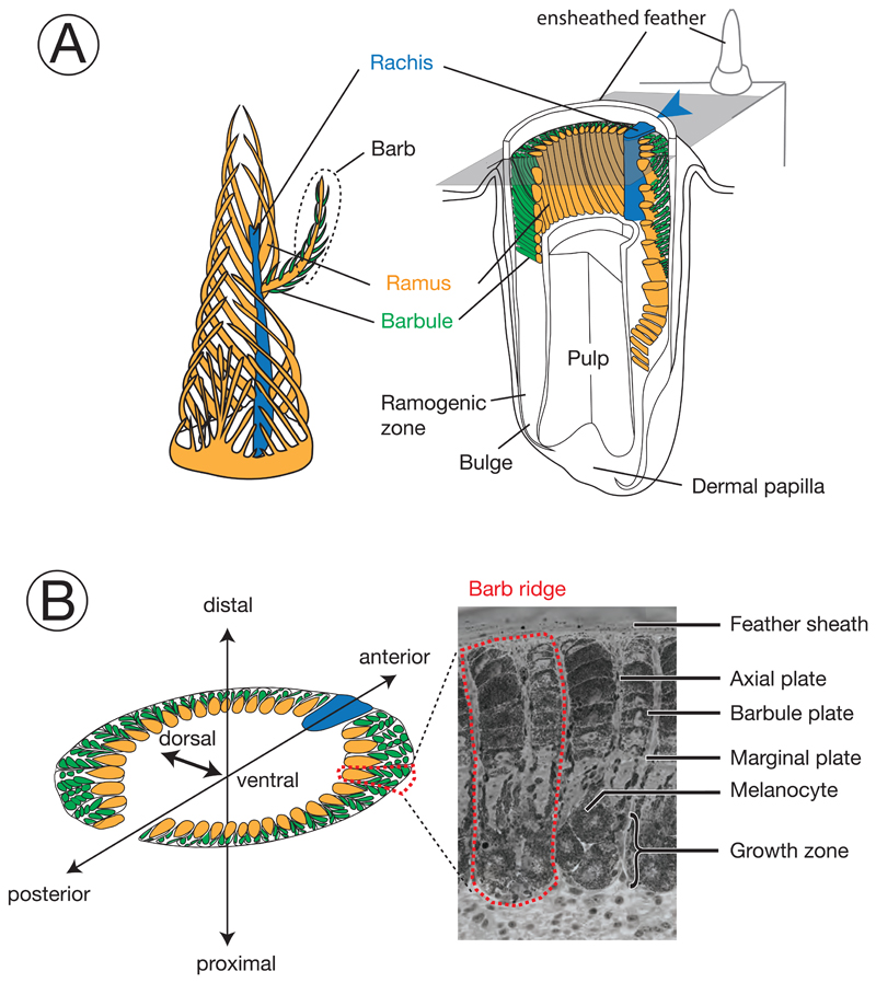
Figure 1. Schematic diagram and terminology of feather follicle morphology.
The feather is a complex epidermal organ of cellular origin. Its basic bauplan follows a hierarchical structure of ramification into barbs and barbules. It develops within follicles, an epidermal modification derived from invagination into the dermis (Chen et al., 2015; d’Ischia et al., 2013). Cells fulfilling different functions in the mature barbs are derived from stem cells at the collar bulge of the basal layer (Yue et al., 2005). Developing progenitors of these cells migrate and gradually differentiate to form mature cells in barbs, including basal cells, marginal plate cells, axial plate cells, and barbulous plate cells (Prum, 1999). Development of the main feather progresses along the proximal-distal axis with helical displacement of barb loci. Barb loci emerge on the posterior midline and are gradually displaced towards the anterior midline where they fuse with the rachis. For more information on the feather morphology and development consult (Lucas and Stettenheim, 1972; Yu et al., 2004).
Colors define homologous structures in the respective panels; arrowheads indicating the rachis facilitate orientation in the remaining figures. A) left: Schematic, three-dimensional representation of a growing pennaceous feather with the afterfeather shown in front. right: Partial section a feather follicle. The cutting plane in grey shows the position of cross sections in panel B. B) left: Schematic cross section of a feather follicle with three-dimensional axes for orientation. right: Example of a cross-section from a growing, ensheathed carrion crow feather displaying two barb ridges.

