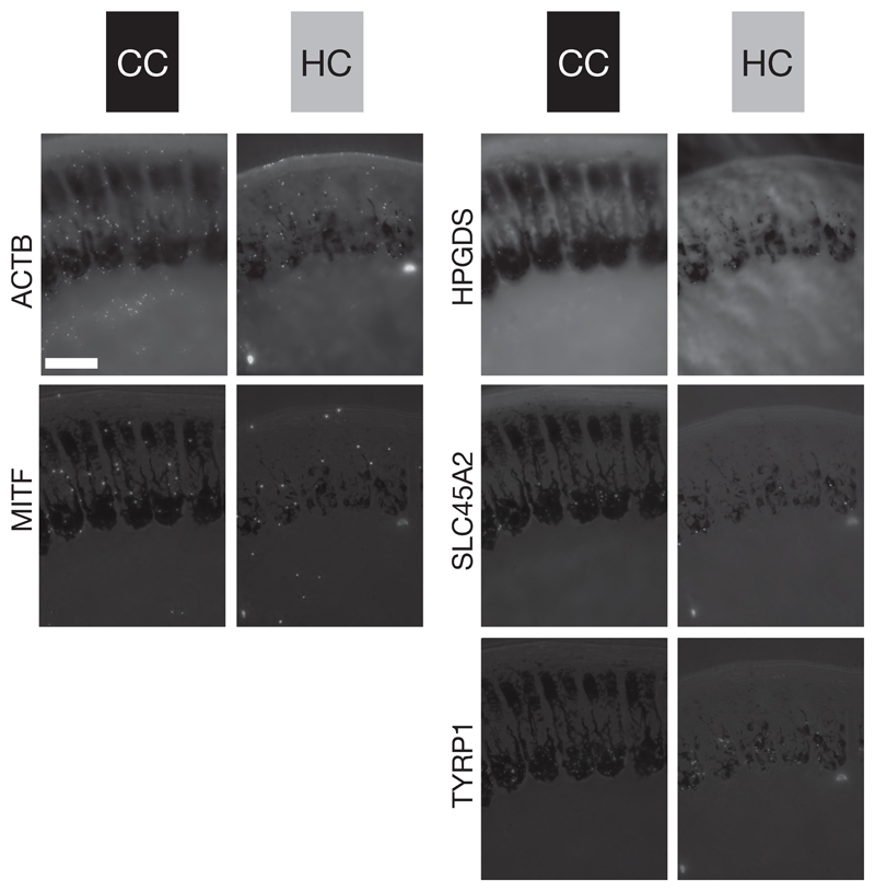
Figure 5. Five-way in situ gene expression in an explicit histological context.
Illustration of in situ stained mRNA transcripts of five genes on the same histological cross section of carrion crow (CC black) and hooded crow (HC grey) follicles sampled from torso at 1000 μm above the dermal papilla. For orientation, these cross sections reflect the schematic shown in Figure 1B. Bright-field images are superimposed for orientation and direct comparison among genes. The images represent one of five serial sections used for statistical analyses. ACTB and MITF (left column) are ubiquitously expressed in all cell types of a follicle. In contrast, HPGDS, SLC45A2 and TYRP1 (right column) are restricted to melanocyte cell bodies; bar = 50 μm.

