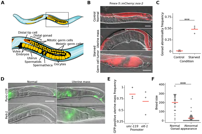
Figure 1. Early-life starvation followed by unrestricted feeding results in reproductive abnormalities.

(A) Cartoon depicting organization of posterior gonad arm of an adult C. elegans hermaphrodite. Boxed area is enlarged to show region assessed for gonad abnormalities. (B) Representative images of control worms and previously starved adults with a germ-cell tumor or uterine mass. DIC and fluorescent images of the germ-cell reporter Pmex-5::H2B::mCherry (naSi2) are overlaid. (C) Frequency of all gonad abnormalities; at least 50 worms per condition per replicate. (D) Representative images of adults after L1 starvation with or without uterine masses. (E) Frequency of GFP-positive uterine masses; at least 20 masses were scored per condition per replicate. (F) Individual brood sizes of worms with either a normal or abnormal gonad; two biological replicates of at least 31 worms each. (B, D) Visible portions of the gonad and uterus are outlined with a white dashed line, animals are oriented as in (A), and scale bar is 50 microns. (C, F) ***p < 0.001; t-test on means of replicates. (C, E) Circles represent biological replicates. (C, E, F) Cross bars reflect the mean. See also Figure S1.
