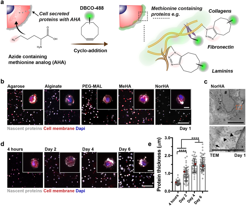
Figure 1. Nascent protein deposition by encapsulated hMSCs occurs early, independent of hydrogel type.
a Schematic of nascent extracellular protein labeling. The methionine analog azidohomoalanine (AHA) is added to the culture media and incorporated into nascent proteins (e.g., fibronectin, collagens, laminins). The bio-orthogonal Cu(I)-free strain-promoted cyclo-addition between the azide and DBCO-modified fluorophore (DBCO-488) enables visualization of the nascent proteins. b Representative images of nascent proteins (white) deposited by hMSCs encapsulated in various hydrogels (alginate, agarose, maleimide modified poly(ethylene glycol) (PEG-MAL), methacrylated hyaluronic acid (MeHA), norbornene modified hyaluronic acid (NorHA)), E = ~9 kPa, scale bar 200 μm, inset 20 μm). c Representative transmission electron microscopy (TEM, * hydrogel, # nucleus; scale bar 5 μm left, 1 μm right) image of encapsulated hMSC after 1 day (24 h in culture). Orange box in top image indicates magnification in bottom image (arrow indicates cell membrane and arrowheads show collagen fibrils). d Representative images of nascent proteins (white) deposited by hMSCs encapsulated in non-degradable NorHA hydrogels (9.0 ± 0.7 kPa, mean ± SD, n = 3 independent measurements) and cultured in growth media (supplemented with AHA) up to 6 days (see Supplementary Figure 3 for daily changes up to 14 days, scale bar 200 μm, inset 20 μm). e Quantification of the accumulated nascent protein thickness deposited by hMSCs encapsulated in non-degradable NorHA hydrogels (n = 40 cells (4 hours), 55 cells (Day 2, Day 4) and 70 cells (Day 6) from 3 biologically independent experiments, mean ± SD, **** p ≤ 0.0001, one-way ANOVA with Bonferroni post hoc, red dots indicate measurements for magnified representative images in d).

