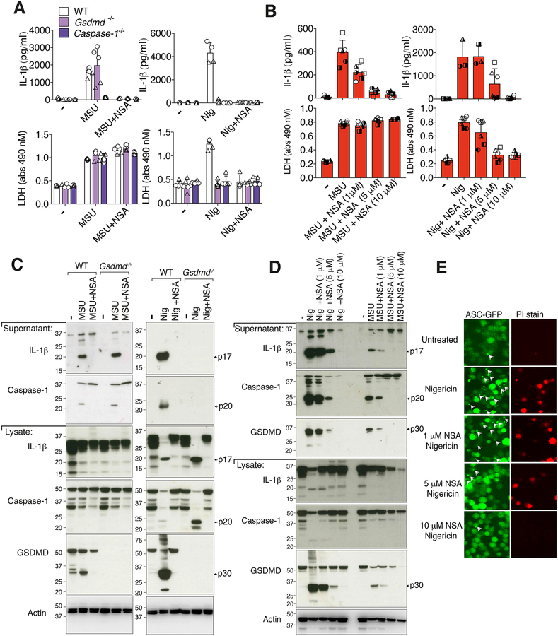
Figure 3. Necrosulfonamide inhibits NLRP3 activation and pyroptosis independent of GSDMD targeting.
(A) IL-1β (top panel) and LDH (bottom panel) levels in the cell supernantant of BMDMs from WT (C57BL/6), Gsdmd−/− and Caspase-1−/− mice primed for 2.5 hr with LPS (50 ng/ml), with necrosulfonamide (NSA) (10 μM) added in the last 30 min of priming, then treated with MSU crystals (300 μg/ml, 6 hr) or nigericin (10 μM, 1 hr). (B) IL-1β and LDH levels in the supernatant of BMDMs from WT (C57BL/6) mice primed with LPS (50 ng/ml, 2.5 hr), with the indicated concentration of NSA added in the last 30 min of priming, followed by treatment with MSU crystals (300 μg/ml, 6 hr) or nigericin (10 μM, 1 hr). (C) WT (C57BL/6) and Gsdmd−/− BMDMs were treated as in (A) and supernatant and total cell lysates analyzed by immunoblot. (D) WT (C57BL/6) BMDMs were treated as in (B) and supernatant and total cell lysates analyzed by immunoblot. Ponceau staining depicts protein loading. (E) Immortalized BMDMs (iBMDMs) expressing ASC-GFP and FLAG-NLRP3 were analyzed by fluorescent microscopy for ASC speck formation (green fluorescence) and PI uptake (red fluorescence) after treatment with the indicated concentrations of NSA for 30–40 min and then nigericin (10 μM, 80 min). ASC specks are indicated with white arrows. (A) Mean ± SD of 4–5 replicates (symbols) pooled from two independent experiments. (B) Mean ± SD of BMDMs from 3–6 mice (symbols) pooled from two independent experiments. (C, D and E) Data are representative of three independent experiments.

