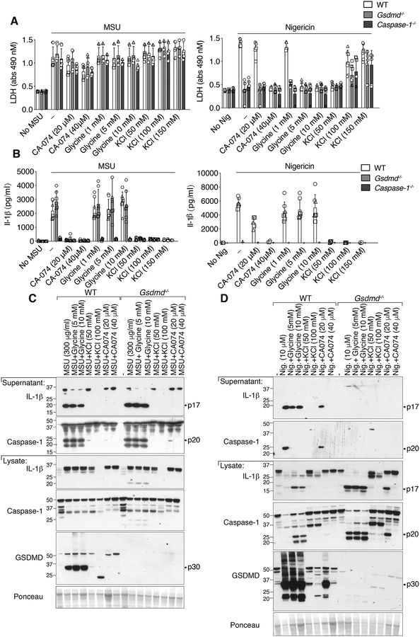
Figure 5. MSU crystal-induced membrane lysis is not significantly reduced by the osmoprotectant glycine, the prevention of potassium ion efflux, or the inhibition of cathepsin activity.
(A) LDH and (B) IL-1β levels in the cell supernatant of BMDMs from WT (C57BL/6), Gsdmd−/− and Caspase-1−/− mice primed with LPS (50 ng/ml, 2.5 hr), with the indicated concentrations of CA-074-Me, glycine and potassium chloride (KCl) added in the last 30 min of priming, and then treated with MSU crystals (300 μg/ml, 6 hr) or nigericin (10 μM, 1 hr). (C and D) BMDMs from WT (C57BL/6) and Gsdmd−/− mice were treated as (A) and cell supernatant and total cell lysates analyzed by immunoblot, as indicated. Ponceau staining depicts protein loading. (A, B) Mean ± SD of 7–9 replicates pooled from three independent experiments. (C, D) Data representative of two independent experiments.

