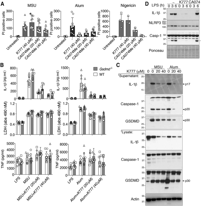
Figure 6. The cathepsin inhibitors, K777 and CA-074-Me, do not prevent MSU crystal-induced death but do reduce inflammasome priming.
(A) BMDMs pre-treated with the indicated cathepsin inhibitors for 40–60 min were then stimulated with MSU (300 μg/ml), alum (300 μg/ml) or nigericin (10 μM) for 4–5 hrs, stained with propidium iodide (PI), and imaged. PI uptake was quantified using Fiji. See Figure S4 for representative images. (B) BMDMs from WT and Gsdmd−/− mice primed with LPS (50 ng/ml, 2.5 hr), and treated in the last 30 min with K777 (20 μM or 40 μM), were then stimulated with MSU (300 μg/ml) or alum (300 μg/ml) for 6 hr. Levels of IL-1β, TNF and LDH in the cell supernatant were measured. (C) BMDMs were treated as in (B) and cell supernatant and total cell lysates analyzed by immunoblot. (D) BMDMs were pre-treated with the indicated cathepsin inhibitor (40 μM) for 40–60 min then stimulated with LPS (100 ng/ml) and total cell lysates examined by immunoblot. *IL-1β band. (A) Mean ± SD of two (nigericin) or three (MSU and alum) pooled independent experiments. Similar symbols represent multiple images per experiment. Different symbols represent independent experiments. (B) Mean ± SD of 6–9 replicates (symbols) pooled from three independent experiments. (C and D) Data are representative of two (C) or three (D) independent experiments.

