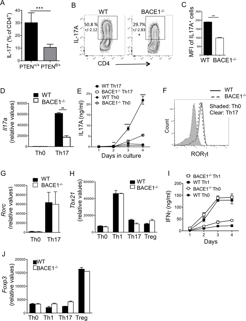
Figure 3: T helper subset differentiation in BACE1−/− T cells.
A: CD4-cre/PTEN+/+ (WT control) and CD4-cre/PTENfl/+ T cells were cultured under Th17 conditions for 3 days and IL-17A+ cells assessed by flow cytometry. B: WT or BACE1−/− CD4+ T cells were differentiated under Th17 conditions and intracellular IL-17A analyzed by flow cytometry on indicated days of culture, mean +/−S.D. indicated. C: Mean fluorescence intensity of IL-17A, gated on IL-17A+ cells, analyzed by flow cytometry on day 3 of culture under Th17 differentiating conditions. D: Gene expression of Il17a in Th0 and Th17 cells on day 3 of culture, normalized to Gapdh. E: IL-17A in culture supernatants analyzed by ELISA at indicated times, IL-17A levels reflect accumulated cytokine from start of culture. F: RORγt protein expression analyzed by flow cytometry on day 3 of indicated cultures. G: Rorc gene expression in T cells cultured under indicated differentiation conditions for two days, normalized to Gapdh. H: Tbx21 (Tbet) gene expression in T cells cultured under indicated differentiation conditions for two days, normalized to Gapdh. I: IFNγ production in Th0 and Th1 cells, analyzed by ELISA at indicated times, IFNγ levels reflect accumulated cytokine from start of culture J: Foxp3 gene expression in T cells cultured under indicated differentiation conditions for two days, normalized to Gapdh. Data indicate mean +/− S.D. of 2–3 replicates representative of at least 4 experiments.

