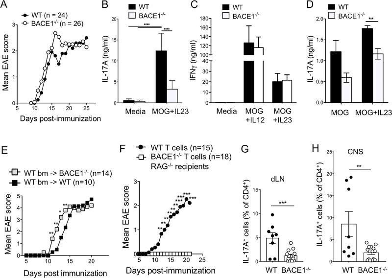
Figure 5: Differential roles for BACE1 in CNS and Th17 cells during EAE.
A: WT and BACE1−/− mice were immunized to induce EAE, and clinical scores monitored, data pooled from 5 experiments. B: Day 8 post-immunization, dLN cells were stimulated with MOG(35–55) + IL-23 for three days then secreted IL-17A analyzed by ELISA. C: Day 8 post-immunization, dLN cells were stimulated with MOG(35–55) + IL-12 or MOG(35–55) + IL-23 for three days, then secreted IFNγ analyzed by ELISA. D: Day 12 post-immunization, dLN cells were stimulated for 18hr with MOG(35–55) +/− IL-23, then secreted IL-17A analyzed by ELISA. B-D data pooled from 3 experiments. E: WT bone marrow was transferred into irradiated WT or BACE1−/− recipients and EAE was induced following 8 weeks reconstitution, data pooled from two experiments. F: WT or BACE1−/− CD4+ T cells were transferred into RAG1−/− recipients, EAE was induced the following day and clinical signs were monitored, data pooled from 3 experiments. G: IL-17A+ cells analyzed by flow cytometry in dLN on day 12 post-EAE induction following PMA/ionomycin stimulation. H: IL-17A+ cells analyzed by flow cytometry in CNS on day 12 post-EAE induction following PMA/ionomycin stimulation. Data in G,H pooled from two experiments, each point represents an individual mouse.

