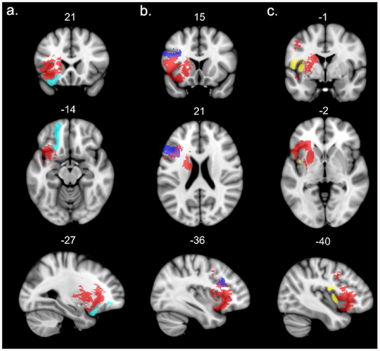
Fig. 4.
Left hemisphere short intralobar association white matter tracts affected by the lesion (probability of disconnection by the VLSM map > 80%). The significant VLSM cluster is shown in red. For the sake of visualization, the probabilistic masks of the tracts are thresholded at minimum 0.7. a – frontal orbito-polar tract, b – frontal inferior longitudinal tract, c – fronto-insular tract4. (For interpretation of the references to colour in this figure legend, the reader is referred to the Web version of this article.)

