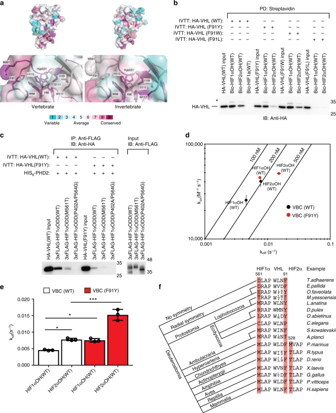
Fig. 5.
VHL Phen+3 is associated with a more stable VHL/HIFα complex. a The structure of HIF1α-VHL complex (PDB: 1LM8) is rendered as a cartoon superimposed with a transparent surface representation. Using multiple sequence alignments for VHL and HIFα in vertebrate and invertebrate species, amino acid residues were color-coded according to conservation using the Consurf webserver. b Biotinylated HIFαOH peptides were immobilized on streptavidin- agarose beads and incubated with in vitro transcribed and translated (IVTT) HA-VHL. Streptavidin beads were pulled down (PD) and levels of HA-VHL were visualized via immunoblotting (IB). c 3xFLAG-HIFα oxygen-dependent degradation (ODD) domains were IVTT and incubated with purified HIS6-PHD2 (181–426). Following hydroxylation (one hour), 3xFLAG-HIFα ODD domain was immobilized on protein A beads coated with anti-FLAG antibody and incubated with IVTT HA-VHL. 3xFLAG-HIFα ODD domain was immunoprecipitated (IP) and levels of HA-tagged VHL were visualized via immunoblotting (IB). Molecular weight markers (kDa) are labeled for western blots. d, e Biolayer interferometry kinetic analysis of VHL-elongin B-elongin C (VBC) complex binding to immobilized HIFα peptides. Biotinylated peptides were immobilized on streptavidin- coated biosensors and binding to VBC complex was monitored. The data was analyzed assuming a 1:1 binding model via the BLItz Pro software. d Rate plane with Isoaffinity Diagonals (RaPID) plot highlighting the kinetic parameters of VBC complex, either WT of F91Y, binding to HIFα peptides. Values represent mean of three experiments conducted with independently purified protein ± s.e.m. e The dissociation constants associated with VBC binding to HIFα peptides are shown on a linear scale. Statistical significance was assessed using a one-way ANOVA with Tukey’s post hoc test. Values represent mean of three experiments conducted with independently purified protein ± s.d. * p < 0.05, *** p < 0.005. f An idealized phylogenetic tree of metazoan evolution is depicted. Short HIF1α, HIF2α, and VHL sequences from representative species are given

