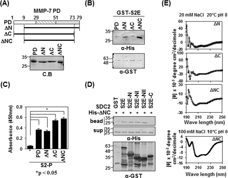
Figure 2.
The pro-domain of MMP-7 is involved in its interaction with the syndecan-2 extracellular domain. (A) Schematic representations of the pro-domain of MMP-7 (PD) and the deletion mutants lacking the pro-domain, N-terminus (ΔN), C-terminus (ΔC) and both N- and C-terminus (ΔNC) (top). His-tagged MMP-7 pro-domain was purified with Ni-NTA agarose beads, separated by SDS-PAGE and stained with Coomassie Blue (bottom). (B) Purified GST-S2E, S2E-C and S2E-NII were incubated with His-tagged PD, or ΔN, ΔC or ΔNC MMP-7 pro-domain. Materials bound to glutathione-agarose beads were immunoblotted with an anti-His tag antibody (top). The membranes were then stripped and re-probed with an anti-GST antibody (bottom). (C) ELISA plates coated with 600 ng of S2-P were incubated with the indicated His-tagged MMP-7 pro-domains (500 ng/well) for 2 h at 20 °C. The plates were washed, incubated with an anti-His tag antibody followed by IgG-HRP, and developed with TMB-ELISA. Absorbance was measured at 450 nm. Data are shown as mean ± S.D. (n = 3), *p < 0.05 versus blank. (D) Purified GST-SDC2 was mixed with His-tagged ΔNC for 2 h at 4 °C., separated by SDS-PAGE, and immunoblotted with anti-His or -GST antibodies. (E) The secondary structures of the MMP-7 pro-domain mutants of ΔN, ΔC and ΔNC were diluted to 50 μM in a buffer consisting of 10 mM HEPES and 20 mM NaCl, pH 8, and kept at 20 °C. The TFE-induced helicity curves were obtained by recording the CD signal for each independent sample from 0% to 40% TFE (100 mM NaCl, pH 6, 20 °C) with far-UV.

