Figure 5.
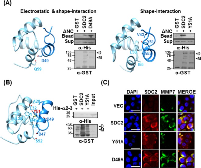
Tyrosine 51 of the syndecan-2 extracellular domain is involved in the interaction of syndecan-2 with the pro-domain of MMP-7. (A) A docking structure for the complex formed between ΔNC (sky blue) and S2-P (dodger blue), as generated by the HEX 6.3 program. Docking states were generated using electrostatic and shape interactions (a), or with shape interactions alone (b). Purified GST-SDC2 or its mutants were incubated with His-ΔNC for 2 h at 4 °C. Materials bound to glutathione-agarose beads were immunoblotted with the indicated antibodies (a or b right). (B) The HADDOCK program was used to generate the docking structure of ΔNC and S2-P. The residues identified as being important matched those identified in our NMR titration experiments. The residues lining the hydrophobic cavity are drawn with a stick model (left). Purified GST-SDC2 and the Y51A mutant were incubated with His-ΔNC for 2 h at 4 °C and the materials that bound to the glutathione-agarose beads were immunoblotted with the indicated antibodies (right). (C) Control HT-29 cells (VEC) and HT-29 cells transfected with vectors encoding SDC2 or mutants were immunostained with anti-syndecan-2 or anti-MMP-7. The results were visualized with Texas Red-conjugated goat anti-rabbit (red) or FITC-conjugated goat anti-mouse (green). DAPI was used to stain nuclei (blue). Scale bar, 20 μm.

