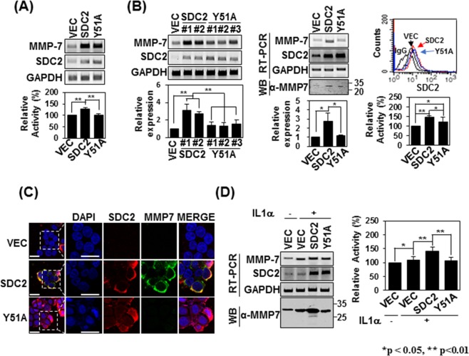
Figure 6.
Tyrosine 51 of the syndecan-2 extracellular domain is involved in regulating pro-MMP-7 activation. (A) HT-29 cells were transiently transfected with 1 µg of vectors encoding SDC2 or the interaction-defective syndecan-2 mutant, Y51A, and the mRNA expression levels of SDC2 and MMP-7 were evaluated by RT-PCR (top). Conditioned media (CM) were collected and proteolytic activity was measured using quenched fluorescence peptide cleavage assay. The relative activity was normalized versus the fluorescence of a vector control (bottom). Data are shown as mean ± S.D. (n = 3), **p < 0.01 versus VEC or SDC2. (B) HT-29 cells were stably transfected with vectors encoding SDC2 or Y51A. The expression levels of the target mRNAs were analyzed by RT-PCR and quantitative real-time PCR (q-PCR) of three independent experiments was performed and normalized to GAPDH expression. Data are shown as mean ± S.D. (n = 3), *p < 0.05, **p < 0.01 versus VEC or SDC2 (left). Flow-cytometric analysis was used to examine membrane-bound SDC2 and Y51A (right top). CM were collected and proteolytic activity was measured using quenched fluorescence peptide cleavage assay (right bottom). (C) Control HT-29 cells (VEC) and HT-29 cells stably expressing SDC2 or Y51A were immunostained with anti-syndecan-2 or anti-MMP-7. The results were visualized with Texas Red-conjugated goat anti-rabbit (red) or FITC-conjugated goat anti-mouse (green). DAPI was used to stain nuclei (blue). Scale bar, 20 μm. (D) The indicated cells were treated with 1 ng/ml of interleukin-1α (IL-1α). The mRNA expression levels of SDC2 and MMP-7 were evaluated with RT-PCR (left top). CM were collected from the indicated cells and immunoblotted with an anti-MMP-7 antibody (left bottom) or subjected to quenched fluorescence peptide cleavage activity assay (right). Data are shown as mean ± S.D. (n = 3), *p < 0.05, **p < 0.01 versus VEC or SDC2.

