Figure 1.
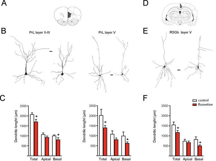
Chronic fluoxetine treatment (28 days) decreased the dendritic length of pyramidal neurons in limbic cortices. Golgi staining was performed to analyze the prelimbic (PrL) and retrosplenial granular b (RSGb) cortices. Neurons were drawn with camera lucida and digitally scanned to visualize the morphology. (A) and (D) The analyzed areas are schematically shown (Paxinos and Watson, 1998). (B) and (E) Representative drawing of pyramidal neurons obtained from vehicle treated (control) or fluoxetine-treated rats. In the PrL cortex, layer II–III and V neurons were analyzed, while in the RSGb cortex, the analysis was restricted to layer V neurons. In each case, the neuron at the left is from the control (vehicle treated), while the neuron at the right is from the fluoxetine-treated animal. Scale bar: 20 µm. (C) and (F) Quantifications of the total, apical, and basal dendritic lengths of neurons in the PrL cortex (layers II/III in the left panel and layer V in the right panel) and RSGb cortices. Values represent the mean ± SEM. For each condition, we analyzed at least 10 neurons obtained from four animals per condition for the PrL cortex and three animals per condition for the RSGb cortex. *p ≤ 0.05, Mann–Whitney U-test.

