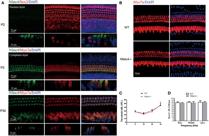
Figure 1.
Nfatc4 was expressed in the cochlear hair cells, and Nfatc4−/− mice showed normal cochlear development and hearing function. (A) Nfatc4 immunofluorescence staining in the cochlear hair cells (middle turns) of P2 and P30 WT mice. Myo7a and Sox2 were used as hair cell and supporting cell markers, respectively. (B) Myo7a immunofluorescence in cochlear hair cells (middle turn) of adult Nfatc4−/− and WT mice. (C) The hearing thresholds of ABR measurement in the adult Nfatc4−/− and WT mice. (D) The numbers of hair cells in Nfatc4−/− and WT mice. Scale bars: 25 μm. n = 5.

