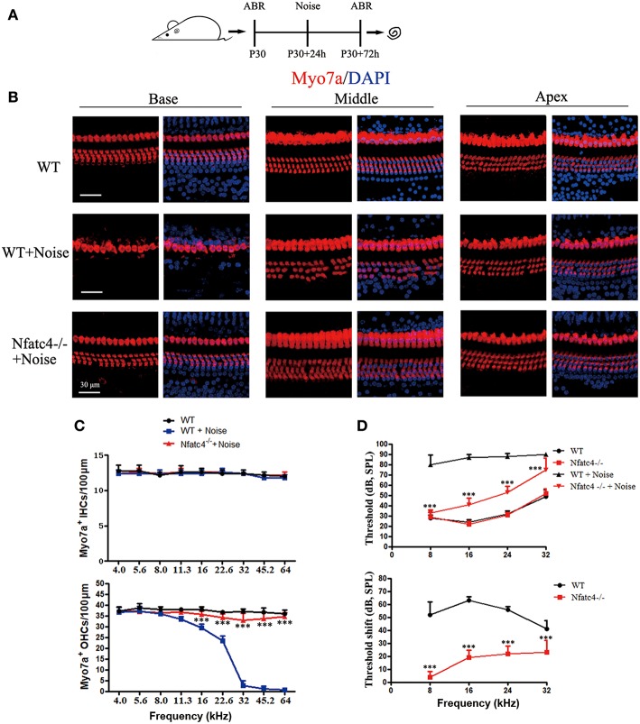
Figure 3.
Nfatc4-deficient mice displayed enhanced resistance to hearing loss induced by noise exposure. (A) The diagram of the assay for B–D. P30 Nfatc4−/− and WT mice (littermates) were examined by ABR, then 24 h later exposed to 118 dB noise (8–16 kHz) for 2 h. At 2 days after noise exposure, hearing function was examined again by ABR and the mouse cochleae were harvested. (B) The representative Myo7a immunofluorescence staining of sensory epithelium from Nfatc4−/− and WT mice after noise exposure. (C) Quantification of inner hair cells (IHCs) and outer hair cells (OHCs). The numbers of OHCs in the middle and basal turns were significantly greater in the cochlear epithelium from Nfatc4−/− mice than in WT mice after noise exposure. (D) Pure-tone ABR thresholds and threshold shift of Nfatc4−/− mice and WT mice after noise exposure. Scale bar = 30 μm. n = 5. ***P < 0.001 vs. the WT+Noise group.

