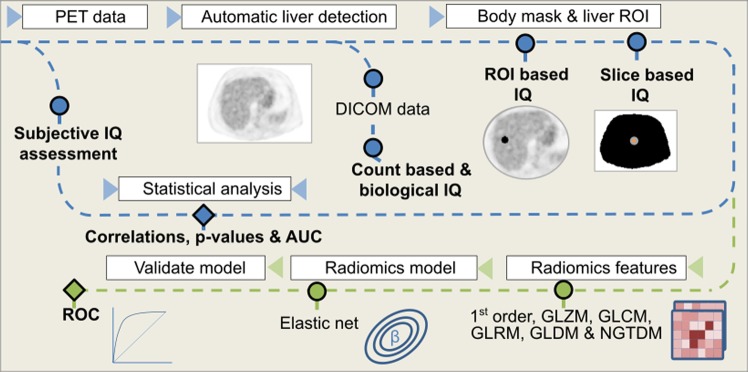
Figure 1.
All image quality features were extracted and processed using an automatic pipeline. Blue line describes the first phase of the methodology: image is converted to SUV units and an automatic algorithm detects the slice including more liver parenchyma. Then, all DICOM data are extracted from the bed corresponding to this slice and a region of interest is placed on the liver to extract ROI-based image quality metrics. From a body mask, all slice-based image quality parameters are extracted. The green line describes the second phase: all common radiomics features are also extracted from the selected slices, as well as from its surrounding volume. Next, an elastic-net model is fitted selecting the relevant features. Results are compared in both lines with the subjective assessment.

