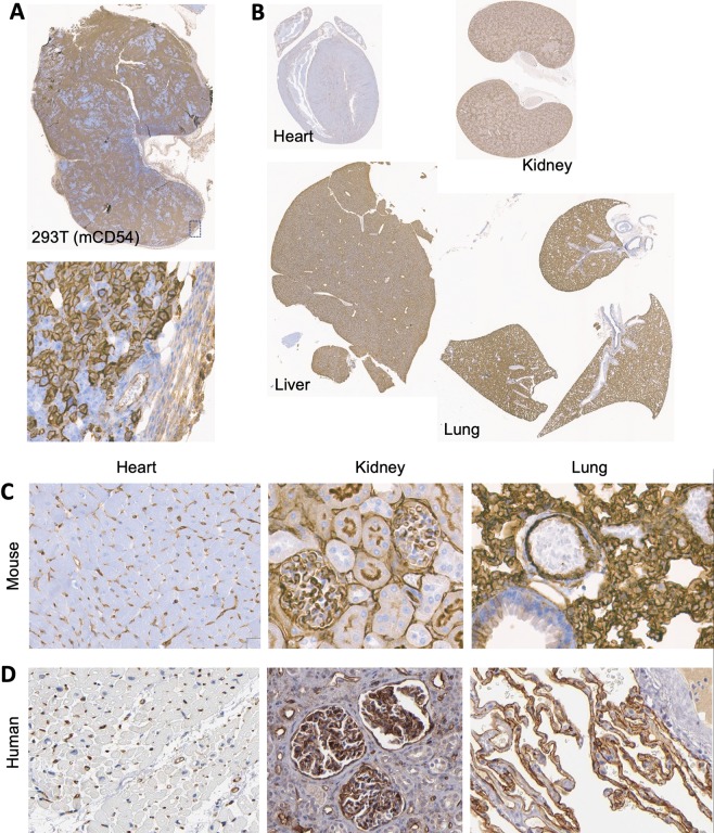
Figure 5.
Comparison of the profile of ICAM-1 expression in tissues of human and mouse. (A) Specificity of anti-mouse ICAM-1 antibody (3E2) by immunostaining was first validated against subcutaneous tumors of 293T cells stably transduced with murine ICAM-1 (mCD54). ICAM-1 staining of the plasma membrane of 293T(mCD54) cells was clearly discerned from little to background staining of stroma cells within and at boundary of tumor. (B) Antibody staining of heart, liver, lungs, and kidneys was shown. (C) ICAM-1 staining was observed in glomeruli endothelial cells of the kidney and alveoli pneumocytes of the lung in mouse tissues. Heart tissue was devoid of specific ICAM-1staining. (D) Similar distribution of ICAM-1 expression in human tissues was observed. ICAM-1 staining of human tissues was obtained from Protein Atlas database (https://www.proteinatlas.org/ENSG00000090339-ICAM1/tissue).

